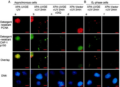
FIG. 4.
In situ visualization PCNA and CAF-1 p150 at SSB. (A) Double immunolabeling for PCNA and CAF-1 p150 in asynchronous cells. Panels in column a are unirradiated XPA-UVDE cells; those in c are DIQ-treated XPA-UVDE cells fixed at 2 min after local UV irradiation (20 J/m2). Those in column b are XPA-UVDE cells fixed at 2 min after local UV irradiation (20 J/m2); those in d are XPA-Vector cells fixed at 2 min after local UV irradiation (20 J/m2). Panels in the uppermost row are cells immunolabeled for PCNA (red), whereas those in the second row are cells immunolabeled for CAF-1 p150 (green); both proteins were in the detergent-resistant form, as described in Results. Colocalization of the proteins appears yellow, as seen in the third row for the overlay of the corresponding panels. The nuclei were stained with DAPI and are shown in the bottom row. Bar, 10 μm. (B) Double immunolabeling for PCNA and CAF-1 p150 in G1-phase cells. XPA-UVDE (column e) and XPA-Vector (column f) cells were treated with mimosine as described in Materials and Methods. Cells were exposed to local UV irradiation (20 J/m2) and incubated for 2 min, then doubly immunolabeled for PCNA (uppermost row, red) and for CAF-1 p150 (second row, green); both proteins were in the detergent-resistant form as described in Results. Colocalization of the proteins appears yellow, as seen in the third row for the overlay of the corresponding panels. The nuclei were stained with DAPI (bottom row). Bar, 10 μm.

