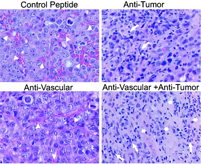
Figure 2.
Histology after combined antiangiogenic and tumor-specific immunotherapy of established primary neuroblastoma tumors, surgically removed 20 days after tumor cell inoculation. Formalin-fixed primary tumors were subjected to paraffin embedding and subsequent hematoxylin/eosin staining. Arrowheads delineate blood vessels. Necrotic areas and leukocyte infiltrates are indicated by open stars and arrows, respectively. Representative areas were photographed at ×630.

