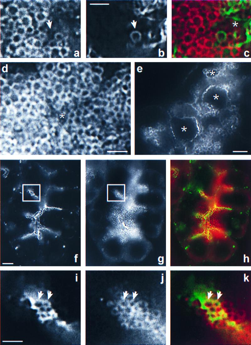
Figure 2.

Rab3D release and actin coating of zymogen granules are interdependent events. (a– c) Double localization of rab3D and filamentous actin in tissue challenged with carbamylcholine (10 μM, 1 h). A close-up of part of an acinar lumen (asterisk) and the underlying zymogen granule field is shown. Rab3D immunofluorescence is depicted in a, and the corresponding phalloidin fluorescence is shown in b. The merge of the rab3D and phalloidin fluorescence images is shown in c. Note that the actin-coated granule (arrow) is not outlined by rab3D immunoreactivity, in contrast to the other zymogen granules. (d) Rab3D immunofluorescence in an acinus of tissue that was stimulated with carbamylcholine, showing absence of rab3D immunoreactivity on the apical membrane surrounding the lumen (asterisk). (e) Rab3D immunofluorescence in an acinus stimulated with carbamylcholine in the presence of cytochalasin D. The dramatically enlarged lumen (asterisks) is now outlined by strong rab3D staining. (f–h) Double localization of rab3D and filamentous actin in an acinus stimulated with carbamylcholine in the presence of latrunculin B and 2,3-butanedione monoxime. Phalloidin fluorescence is shown in (f). Note the presence of numerous actin-coated granules packed in clusters. Also note that the acinar lumen has not expanded. The corresponding rab3D immunofluorescence is depicted in g, and the merge between both fluorescence images is shown in h. Boxes in f and g outline an area that is shown at higher magnification in i (phalloidin), j (rab3D), and k (merge). Note that the actin-coated granules (two are indicated by arrows) are rab3D-immunoreactive. [Bars = 2 μm (b, d, and i) and 5 μm (e and f).]
