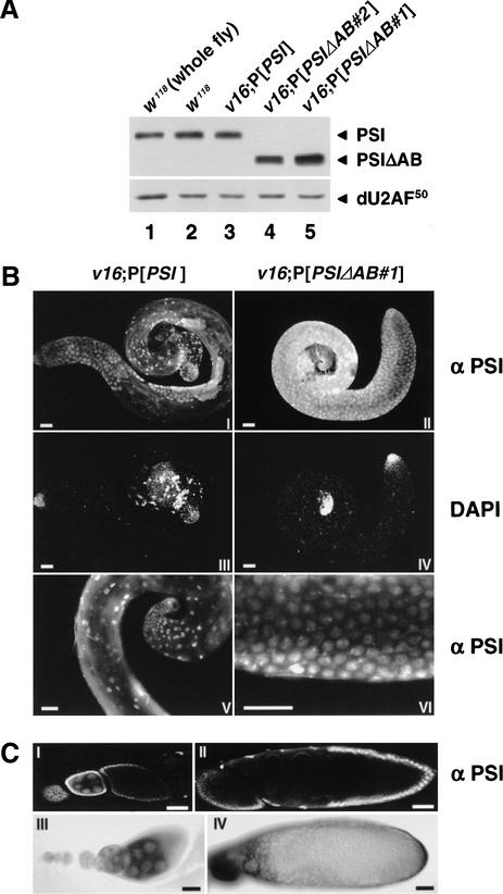
Figure 6.
Pattern of PSI expression in Drosophila testes and ovaries. (A) Immunoblot analyses of testes protein extracts prepared from w1118 males (lane 2) or homozygous v16 males rescued by the PSI (lane 3) or PSIΔAB (lanes 4,5) cDNA transgenes were performed as in Figure 1B. A w1118 whole-fly protein extract was processed in parallel (lane 1). (B) Testes dissected from homozygous v16 adult males rescued by the PSI (I,III,V) or PSIΔAB#1 (II,IV,VI) cDNA transgenes were fixed and stained with affinity-purified anti-PSI rabbit antibodies and Alexa Fluor secondary antibodies (I,II,V,VI) and with DAPI (III,IV). Magnification of the basal end of a v16;P[PSI] testis (V) or the central part of a v16;P[PSIΔAB#1] testis (VI) shows the nuclear localization of both the PSI and PSIΔAB proteins in somatic and germ-line cells. Scale bar, 50 μm. (C) Stage 4–9 (I), 2–8 (III), 10 (II), or mature (IV) oocytes from w1118 females were fixed and stained with anti-PSI polyclonal antibodies (I,II) or hybridized with a digoxygenin-labeled anti-sense PSI RNA probe (III,IV). To observe the very low level of PSI protein expression, whole-mount ovarioles were analyzed by confocal imaging. Scale bar, 50 μm.

