
Figure 6.
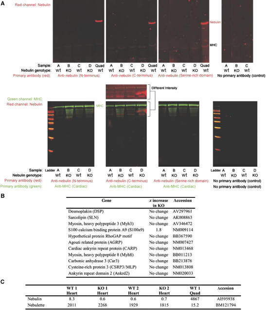
Protein and transcript studies in myocardium of NEB KO mice. (A) Two-color Western blots of NEB KO and wt mouse cardiac muscles. Left ventricle muscles from four mice were tested with three different nebulin antibodies. The four samples under investigation were (lanes marked A) 10-day-old wt, (lanes marked B) 10-day-old NEB KO, (lanes marked C) 5-day-old wt, and (lanes marked D) 5-day-old NEB KO. Positive controls were quadriceps (Quad) from a nebulin wt mouse and an antibody to MHC. Top: Screen of nebulin in the molecular mass region between titin and MHC. Bottom: Proteins smaller than MHC were also tested with nebulin antibodies (Mr standards from small to large: 20, 30, 40, 50, 60, 80, 100, and 120 kDa). A separate view of the intensified red channel (anti-nebulin C-terminus) is also shown. This reveals that weak signals are detected with the anti-nebulin antibody but that they are not differential between wt and KO lanes. (B) Gene expression profiling in cardiac muscle at day 14 identified no or minimal changes. Shown are 11 of the genes which were highly upregulated in skeletal muscle of nebulin deficient mice (see Figure 3). (C) Array data (absolute signal strength) show the de facto absence of nebulin transcripts in wt and KO heart for the exons contained on affymetrix array (Mouse Genome 430 2.0). Nebulette is present in wt heart but reveals no differences between wt and KO animals. For comparison the absolute magnitude of skeletal muscle signals is shown.