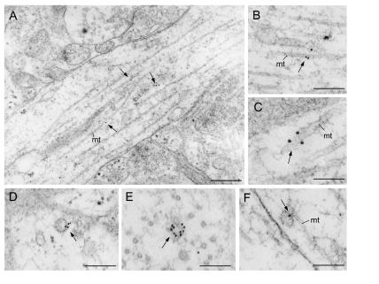
Figure 5.
Immunogold labeling of hippocampal organotypic cultures for mVAP33. Immunoreactivity was mainly associated with microtubules (A), where it was often clustered (B and C). Clustered labeling was also associated with the membranes of vesicular structures (D and E). Gold particles were also occasionally seen between a vesicle and a microtubule (F). There was no detectable signal associated with synaptic vesicles. (Bars: A, 300 nm; B and D, 200 nm; C, 100 nm; E, 150 nm; and F, 80 nm.)

