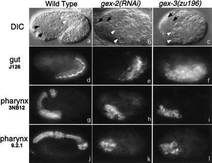
Figure 2.
gex mutant embryos fail to undergo morphogenesis but do differentiate specific tissues. (Top panels) Nomarski images. All other panels are fluorescent images using monoclonal antibodies to pharynx (3NB12 and 9.2.1) and intestine (J126). Anterior is left, and dorsal is up. Wild-type N2 embryos at this stage have the internal organs of the pharynx and intestine fully enclosed by the hypodermis. Black-tipped arrows point to the anterior of the pharynx, and white-tipped arrows point to the anterior of the intestine. Antibodies 9.2.1 and J126 illustrate that the pharynx and intestine form elongated tubular structures. In gex-2 and gex-3 embryos the hypodermis never encloses the embryo and instead constricts on the dorsal side. Embryos end embryogenesis with internal organs that lie on the outside and do not form the correct elongated tubular structures.

