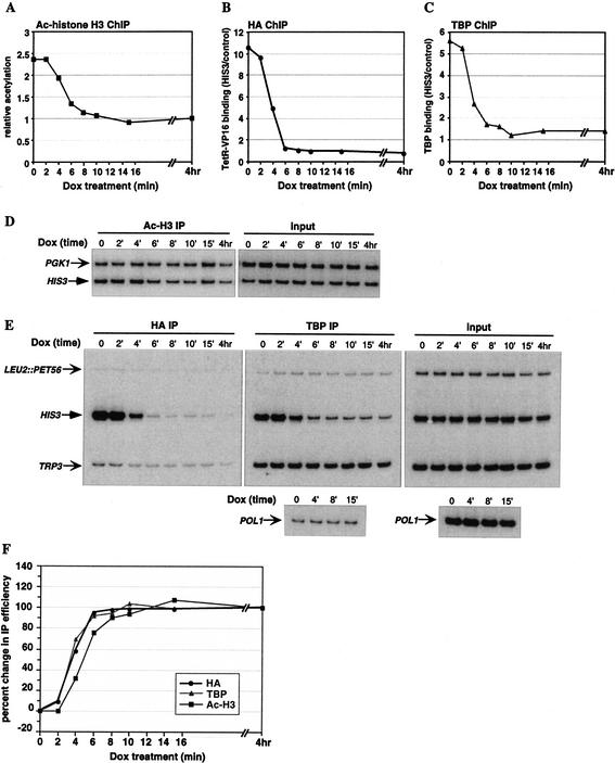
Figure 4.
Reversal of targeted histone acetylation and TBP binding upon dissociation of TetR–VP16 from the HIS3 promoter. Cross-linked chromatin from TetR–VP16-expressing YKT2 cells (contain a nonactivated HIS3 promoter) treated with Dox for 0–15 min or 4 h was immunoprecipitated with antibodies to acetylated histone H3, the HA epitope for measuring TetR–VP16 occupancy, and TBP, and analyzed by quantitative duplex PCR. (A,D) Histone acetylation at the nucleosome +2 region of HIS3 was analyzed as described in Figure 3. (B,E) TetR–VP16 binding to the HIS3 promoter was determined by normalizing each IP signal to its corresponding input signal, and dividing the HIS3 results by the control TRP3 results. (C,E) TBP binding to the HIS3 promoter was determined by normalizing each IP signal to its corresponding input signal, and dividing the HIS3 results by the control TRP3 results. The POLI ORF signal represented background binding by TBP, and the results are expressed relative to the average normalized POL1 background signal. (F) Kinetics of changes in histone acetylation, and occupancy by TetR–VP16 and TBP at the HIS3 promoter throughout the time course. At each time point, the change in IP value from the time 0 value was determined, and expressed relative to the change at 4 h set as 100%.

