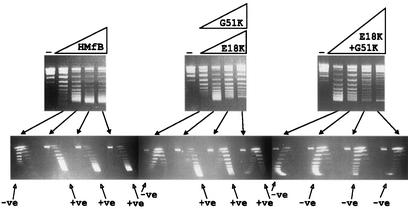
FIG. 3.
One- and two-dimensional agarose gel electrophoretic separations of the topoisomers generated by HMfB or HMfB variants assembled on relaxed, circular pUC18 DNA molecules. The procedures used to assemble complexes, remove plectonemic supercoils, deproteinize, and separate pUC18 topoisomers by one- and two-dimensional agarose gel electrophoresis have been described previously (9, 10). The control lanes (−) contained relaxed pUC18 DNA; adjacent lanes contained the pUC18 topoisomers generated by HMfB or HMfB variant assembly on aliquots of this DNA at histone-to-DNA mass ratios of 0.4, 0.6, 0.8, and 1. Almost identical results were obtained with the E18K variant (shown) and G51 variant. Topoisomers were separated in the first dimension (upper gels) on the basis of linking number, with increasing linking number resulting in increasing mobility, and in the second dimension (lower gels) on the basis of negative (−ve) or positive (+ve) supercoiling. Nicked and relaxed circular DNAs migrated together and formed the band near the top of each gel.

