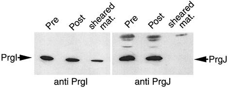
FIG. 4.
Analysis of the needle substructure protein content. Cultures of wild-type bacteria were grown under inducing conditions and pelleted by low-speed centrifugation (10,000 × g). Pellets were washed once with phosphate-buffered saline (PBS), repelleted, and resuspended in PBS. Cells were then passed through a 20-gauge needle 20 times in order to shear off the needle portion of the needle complex. Cells were pelleted and the supernatant was removed and subjected to TCA precipitation. The cell pellet was lysed as described for Fig. 3. Lysate pellets of both unsheared control cells and the sheared cells along with the pellet from the TCA-precipitated supernatant were analyzed by immunoblotting using anti-PrgI or anti-PrgJ antibodies as described above.

