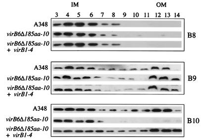
FIG. 6.
Membrane localization of VirB8, VirB9, and VirB10 from A348, A136(pAB123, pZL48-D5, pZL3) (virB6Δ185aa-10, virB1 to virB4) and A136(pAB123, pZL48-D5) (virB6Δ185aa-10). Membrane fractions were separated by sucrose density gradient as described in Materials and Methods. Membrane fractions 3 to 14 (from the top to the bottom of the gradient) were subjected to SDS-PAGE, blotted, and probed with anti-VirB8, anti-VirB9, or anti-VirB10. Similar results were observed in three other experiments. IM, inner membrane; OM, outer membrane.

