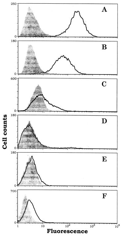
Figure 3.
FACS assays for binding of the immunoconjugate G71–1 to human melanoma cells and human control cells. (A) Melanoma lines, TF2. (B) Melanoma line A-2058. (C) Melanocytes. (D) Microvascular endothelial cells. (E) Fibroblast cells. (F) Transformed kidney line 293-EBNA. The cells were collected from a culture flask after detachment in a nonenzymatic dissociation medium (Sigma). The detached cells were either exposed to the immunoconjugate (outlined curve) or were not exposed (shaded curve). An increase in fluorescence after exposure to the immunoconjugate indicates that the cells bind the immunoconjugate. The number of gated events analyzed for each sample was at least 5,000. The immunoconjugate E26–1 also was analyzed by FACS and showed similar results as G71–1.

