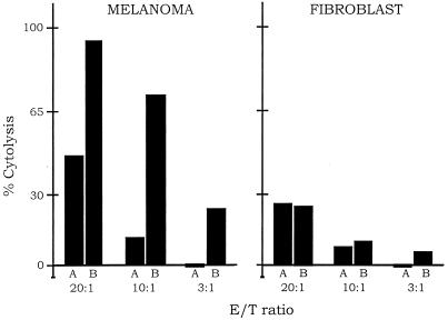
Figure 5.
Immunoconjugate-dependent lysis of melanoma cells by NK cells. The melanoma cell line A-2058 and the fibroblast cell control were labeled with the fluorescent dye Calcein-AM. The fraction of melanoma or fibroblast cells remaining intact after exposure to NK cells alone (A bars), or to NK cells with the immunoconjugate E26–1 (B bars), was measured by residual fluorescence. The ratio of NK effector cells to target cells (E/T) was varied from 3 to 20. Three complete sets of experiments were done for both the melanoma and fibroblast cells; for each experiment, the cytolysis assays were done in quadruplicate. The bars represent the average of the cytolysis assays for the three experiments, which generally agreed within 10%. The % cytolysis was calculated as described in Materials and Methods. Similar results were obtained with the immunoconjugate G71–1.

