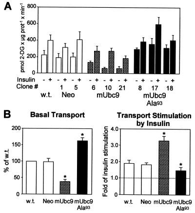
Figure 3.
Modulation of glucose transport by mUbc9. (A) Basal and insulin-stimulated glucose transport rates in wild-type (w.t.), Neo, mUbc9, and mUbc9-Ala93 myoblasts. Cells cultured in 35-mm diameter wells were serum-starved overnight and then incubated in the presence or absence of 1 μM insulin for 30 min. Transport was started by adding 1 μCi/ml 2-[3H]deoxy-d-glucose (NEN) to a concentration of 50 μM for 10 min at 20°C. (B) Basal glucose transport (Left) and the fold stimulation of glucose transport by insulin (Right) in nontransfected (w.t.), Neo, mUbc9, and mUbc9-Ala93 L6 myoblasts. The results represent mean values of four independent experiments performed on individual L6 clones. *, P < 0.05 vs. Neo and wild type by unpaired Student's t tests.

