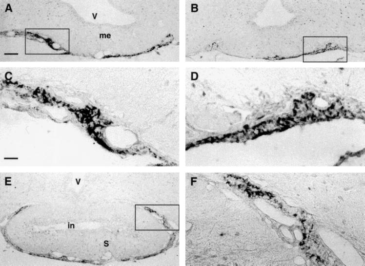
Figure 3.
(A and B) Panoramic overview of two neighboring sections of juxtaneural PT region to compare the distribution of guanylin immunoreactivities obtained with the antisera K605 (A) and K42 (B). (C and D) Higher magnification of boxed areas in A and B, respectively. The immunoreactive elements showed a stick- or rod-like morphology. (E) Guanylin immunoreactive (K605) elements in PT at the pituitary stalk level. (F) Higher magnification of the boxed area in E. V, third ventricle; me, median eminence; in, infundibular recess; S, pituitary stalk. (A, B, and E, bar = 100 μm; C, D, and F, bar = 25 μm.)

