Figure 5.
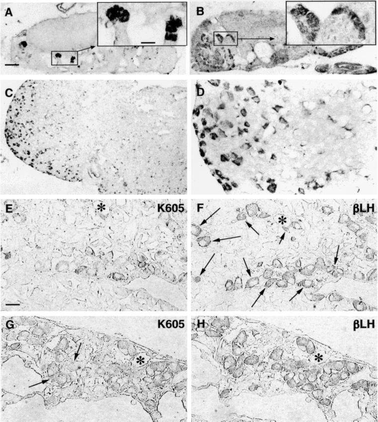
(A and B) Clusters of guanylin immunoreactive cells can be observed in two distal PT sections at different craniocaudal level. (Insets) Higher magnification of boxed areas. (C) Guanylin immunoreactive cells in PD assumed the morphology of the endocrine cells of this region. (D) Higher magnification of an area in C. (E–H) Pair of two consecutive semithin (0.5-μm) sections of PD immunostained for guanylin (K605; E and G) and h-β-LH (βLH; F and H). Almost all guanylin immunoreactive cells show coincident immunoreactivity for h-β-LH, but the number of colocalizing gonadotrophs varies: about one-third in E and F (arrows) and almost all in G and H. Arrows in G indicate the only two guanylin unreactive gonadotrophs. Asterisks label some landmarks to allow the alignment of consecutive sections. (A–C, bar = 100 μm; Insets and E–H, bar = 25 μm.)

