
FIG. 6.
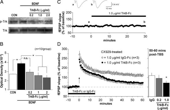
Rescue of LTP by ampakine pretreatment is reversed by the BDNF scavenger TrkB-Fc. A and B: quantitative analysis of phosphorylated Trk protein (pTrk) levels in adult hippocampal slices was used to assess an effective dose range for TrkB-Fc, a scavenger of extracellular BDNF. A: Western blots showing (top blot) that slices receiving 30-min infusions of 60 ng/ml BDNF had greater pTrk levels than did untreated control (CON) slices, and that pTrk levels were reduced by TrkB-Fc in a dose-dependent fashion (right 3 lanes show samples from slices treated with 60 ng/ml BDNF plus the TrkB-Fc dose indicated in μg/ml). Treatments had no effect on total Trk protein levels (bottom blot). B: plot shows mean (±SE) pTrk band densities for 10 Western blot experiments of the type described in A (*P < 0.05; n.s., not significant). C: effects of TrkB-Fc (1.0 μg/ml; black bar) on baseline synaptic responses were assessed using Schaffer-commissural evoked potentials recorded in CA1 str. oriens. This plot, and representative fEPSP traces (inset), indicate that TrkB-Fc infusion did not alter responses to single-pulse stimulation. D: TBS was delivered (time 0, arrow) to slices from CX929-treated rats that had been infused with either 1.0 μg/ml TrkB-Fc (closed circles) or 1 μg/ml IgG-Fc (open circles) for 30 min (black bar); the plot summarizes mean (± SE) values for fEPSP slopes. As shown, TrkB-Fc blocked the stabilization of LTP leading to a slow decay in potentiation, whereas infusion of the control IgG-Fc had no effect. Bar graph to the right describes the percentage potentiation averaged for minutes 50-60 post-TBS for slices from ampakine-treated rats infused with 1 μg/ml IgG-Fc, 0.2 μg/ml TrkB-Fc, or 1 μg/ml TrkB-Fc (n = 3/group for all plots; *P < 0.05 for comparison to IgG group).