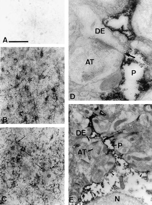
Figure 3.
Expression of NPC1 in monkey temporal cortex by light (A–C) and electron microscopic (D and E) peroxidase immunocytochemistry. (A) Control section incubated with antigen-absorbed NPC1-C antibody showing complete absence of staining. (B) Layer three of cortex showing NPC1 immunoreactivity localized predominantly in processes in the neuropil (*). (C) Higher magnification of layer three showing unlabeled cell bodies (*) surrounded by large numbers of NPC1 positive processes (arrows). (D) Astrocytic process (P) in the neuropil. These processes are present at the sides of asymmetrical synapses (arrows in D and E) between unlabeled axon terminals (AT) and dendrites (DE). (E) Same process (P) as in D, on the side of an asymmetrical synapse (arrow) between an unlabeled axon terminal (AT) and dendrite (DE). The process can be traced to its parent cell body. Label is present in the process and near the cell membrane of the soma (arrowheads), but is absent from the nucleus (N) and more central regions of the cytoplasm. [Bars = 16 μm (A and C), 80 μm (B), 0.3 μm (D), and 1 μm (E).]

