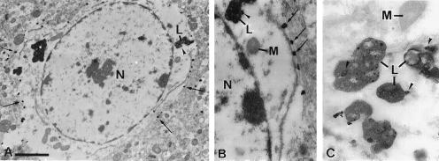
Figure 4.
Electron micrographs showing the distribution of NPC1 immunoreactivity by immunogold/silver labeling in monkey frontal cortex. (A) Cell body of an astrocyte showing gold/silver complexes at the periphery of the cell (arrows) and in association with a lysosome (L, arrowhead). Label is absent from the nucleus (N) and the perinuclear cytoplasm. (B) Higher magnification of A showing a punctate pattern of gold/silver complexes near the cell membrane (arrows) and on the lysosome (L, arrowhead). The nucleus (N) and mitochondria (M) are unlabeled. (C) Gold-labeled lysosomes (L, arrowheads) and unlabeled mitochondrion (M) from another astrocyte. [Bars = 1.8 μm (A) and 0.7 μm (B and C).]

