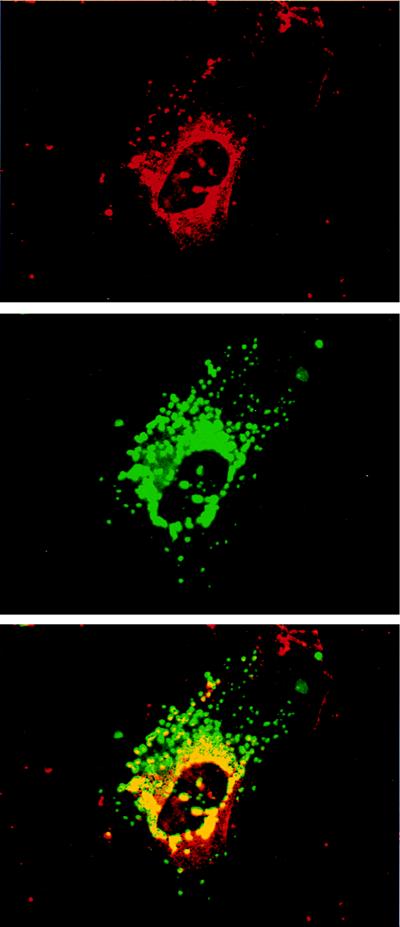
Figure 5.
Confocal microscopic images of cultured human astrocytes showing localization of NPC1 by rhodamine fluorescence (red), LAMP2 by fluorescein fluorescence (green), and colocalization of the two antigens (yellow/orange). NPC1 is present in perinuclear cytoplasmic vesicles. LAMP2 localizes to cytoplasmic vesicles distributed around the nucleus as well as in the periphery of the cell (Middle). Overlapping the images reveals colocalization of NPC1 within the cores of LAMP2 positive vesicles (Bottom).

