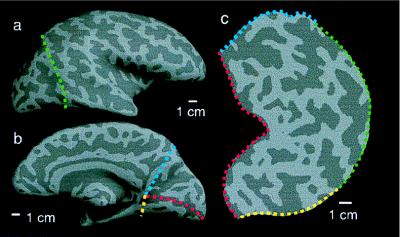
Figure 1.
Cortical reconstruction and flattening. (a) Lateral and (b) medial views of a mathematically inflated cortical surface reveal buried sulci (gyri, light gray; sulci, dark gray). The posterior portion is cut off (green, yellow, and blue lines) and cut along the fundus of the calcarine sulcus (red line in b). (c) The resulting cortical patch is unfurled and laid flat for data visualization.

