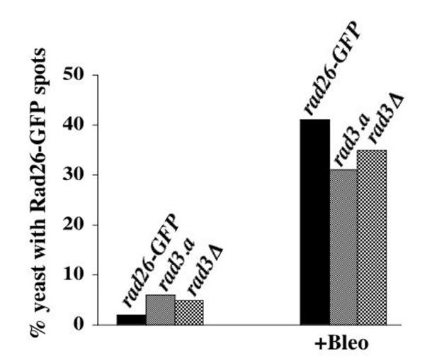
Figure 8.
Rad26-GFP spots form in Bleomycin-treated rad3.a and rad3Δ cells. Cultures were grown at 30°C in liquid, complete media to O.D. 0.3 and then treated with 5 mU/ml of Bleomycin for 3 hours. Cells were then prepared for microscopy following the Triton X-100 extraction method (see Methods). This experiment was repeated three times with similar results, one of which is shown. rad26-GFP (TE1197), rad3.a rad26-GFP (TE1195) and rad3Δ rad26-GFP (1191)

