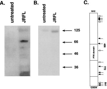
Figure 6.
HIV envelope-induced FAK fragments consistent with cleavage by caspase-3 and caspase-6. An anti-FAK Western blot of untreated or HIV-1 JRFL envelope-treated CD4+ T cell lysates (6 × 10⁷ cells each lane) followed by immunoprecipitation with an anti-phosphotyrosine mAb is shown in A. (B) Immunoprecipitates were stained with anti-PYK2 antiserum. (C) A schematic of all reported caspase-3 and caspase-6 cleavage sites within FAK.

