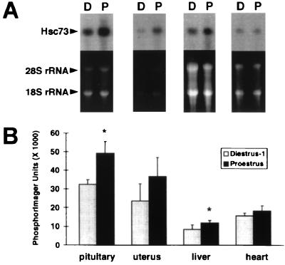
Figure 3.
Northern analysis of Hsc73 mRNA in several peripheral tissues of rats at proestrus and diestrus-1. (A) Total RNA from pituitary, uterus, liver, and heart was subjected to Northern analysis with a Hsc73 cDNA probe prepared from plasmid pP-U.29. Hsc73 hybridization signals from representative animals at diestrus-1 and proestrus are shown above the corresponding ethidium bromide-stained gels. The intensely staining 28S and 18S rRNA bands are indicated. (B) To quantitate the Northern results, the intensity of the Hsc73 hybridization signal, determined by PhosphorImager analysis, was normalized to the amount of RNA loaded in the corresponding lane of the gel. Each bar represents the mean ± SEM. ∗ indicates a significant difference (P < 0.05) between animals at proestrus (n = 3) and diestrus-1 (n = 4). The magnitude of enhancement of Hsc73 mRNA at proestrus relative to diestrus-1 observed in pituitary, uterus, and liver was 52%, 55%, and 40%, respectively.

