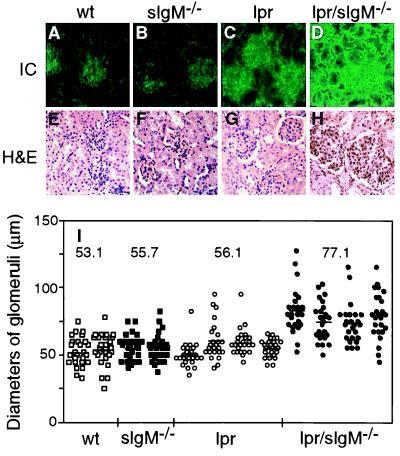
Figure 3.
Lpr/sIgM−/− mice developed more severe glomerulonephritis. (A–D) Representative immunofluorescence stains for IgG-containing immune complexes in glomeruli of wild-type (wt), sIgM−/−, lpr, and lpr/sIgM−/− females at 3 months of age. Kidney sections were stained with a FITC-labeled goat anti-mouse IgG Ab (×40). (E–H) Representative histological stains of kidney sections of wt, sIgM−/−, lpr, and lpr/sIgM−/− females at 6 months of age. Kidney sections were stained with hematoxylin/eosin (×40). (I) Comparison of the diameters of glomeruli in wt, sIgM−/−, lpr, and lpr/sIgM−/− females at 6 months of age. The diameters of 25 randomly selected glomeruli were measured for two wt mice, two sIgM−/− mice, four lpr mice, and four lpr/sIgM−/− mice. Because glomeruli were not perfectly round, each given diameter was an average of the smallest and largest measurements. Each dot represents one glomerulus. The numbers represent the average diameters of glomeruli of specific genotypes.

