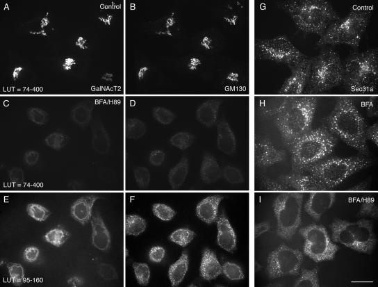
Figure 1.
BFA/H89 disperses GalNAcT2-GFP and GM130 into ER-like structures with extensive ER exit site breakdown. HeLa cells stably expressing GalNAcT2-GFP were left untreated (A, B, and G), treated with BFA for 30 min supplemented with H89 for the last 10 min of BFA treatment (C–F and I), or treated with BFA for 30 min at 37°C (H). The localization of GalNAcT2-GFP was determined, and cells were stained for GM130 or Sec31a (ER exit marker) by immunofluorescence. All GalNAcT2 images were taken at the same exposure conditions. Similarly the GM130 images were taken at constant exposure conditions. Images as indicated are displayed to different LUT (lookup table). All micrographs were taken with a CARV I spinning disk confocal device. Bar, 10 μm.

