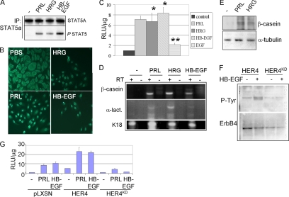
Figure 1.
ErbB4 ligands activate STAT5A in HC11 cells. (A) STAT5A immunoprecipitates (IP) from HC11 cells cultured in serum-free (SF) media with PRL, HRG, or HB-EGF for 30 min were analyzed by immunoblot (IB) with indicated antibodies at the right of each panel. (B) Immunohistochemistry detecting STAT5A localization in serum-starved HC11 cells treated with indicated factors for 30 min. Scale bar, 100 μm. (C) HC11 cells transfected with the STAT5A-responsive promoter, pβcasein-lux, and cultured 48 h in SF media. Agonists were added as indicated for the final 24 h of the experiment. Luciferase activity is shown as the average relative light units (RLU) per μg of total protein ± SD. Experiments were repeated three times, with each sample being analyzed in triplicate. *p < 0.003 versus control. **p < 0.01 versus control. Student’s unpaired t test. (D and E) Confluent HC11 cells cultured in priming media for 2 d and then in priming media ± PRL, HRG, or HB-EGF (where indicated) for an additional 2 d. (D) RT-PCR analysis used primer sets to detect expression of β-casein, α-lactalbumin (α-lact.), or keratin 18 (K18). For each sample analyzed, the reverse transcriptase (RT) was either included (+) or left out (−) of the reaction. (E) Whole cell extracts were analyzed by Western analysis using antibodies against β-casein and α-tubulin. (F) HC11 cells were stably transfected with a pLXSN-derived construct encoding human ErbB4/HER4 or a variant harboring a point mutation at lysine 751 [rendering the product kinase-dead (HER4KD)]. Pooled clones of HC11-HER4 and HC11-HER4KD cells were analyzed by Western blot to detect tyrosine phosphorylation of ErbB4 immunoprecipitates. Serum-starved cells were treated with HB-EGF for 10 min. Blots were stripped and reprobed with a mAb against ErbB4. (G) Pooled clones of HC11-pLXSN, HC11-HER4, or HC11-HER4KD cells were transfected with pβcasein-lux and then treated with PRL or HB-EGF for 24 h, measuring luciferase activity as described above.

