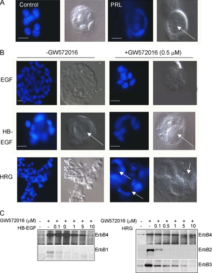
Figure 2.
ErbB4 activity is required for 3D lumen formation. (A) 3D culture of HC11 cells in growth factor-reduced Matrigel ± PRL. Cells were cultured 10 d, stained with DAPI, and photographed using a Zeiss LCM-210; DAPI and DIC images are shown. Arrows indicate lumen formation. Scale bars, 25 μm. (B) 3D culture of HC11 cells in growth factor-reduced Matrigel ± EGF, HB-EGF, or HRG, and ±GW572016 (0.5 μM). Cells were cultured 10 d, stained with DAPI, and photographed using a Zeiss LCM-210; DAPI and DIC images are shown. Arrows indicate lumen formation. Scale bars, 25 μm. (C) HC11 cells were serum/EGF-starved in the presence or absence of increasing concentrations of GW572016 (0–10 μM) for 16 h, and treated for 30 min with HB-EGF or HRG. ErbB1, ErbB2, ErbB3, and ErbB4 immunoprecipitates (IPs) were analyzed by immunoblot for phosphotyrosine.

