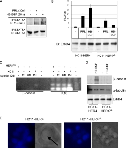
Figure 7.
Cleavage of ErbB4 at V675 is required for ErbB4-mediated differentiation. (A) Pooled clones of HC11-HER4VA cells were serum-starved overnight and then treated for 30 min with PRL or HB-EGF. STAT5A immunoprecipitates (IP) were analyzed by immunoblot (IB) to detect phospho-STAT5A/B or STAT5A. (B) HC11-HER4 and HC11-HER4VA cells were transiently transfected with pβcasein-lux. Cells remained in serum-free media or were treated with the indicated factors for the final 24 h of the experiment. Luciferase activity was determined in extracts as described above (top panel). Extracts were further examined for total levels of HER4/ErbB4 expression by immunoblot using a monoclonal ErbB4 antibody (bottom panel). (C) RT-PCR analysis to detect expression of β-casein or K18 in HC11 or HC11-HER4VA cells treated with PRL or HB-EGF (HB). (D) Western analysis to detect expression of total HER4 levels and β-casein or α-tubulin in extracts from HC11-HER4 and HC11-HER4VA cells cultured in priming media for 48 h and then in priming media ± HB-EGF for an additional 48 h. (E) 3D culture of HC11-HER4 or HC11-HER4VA cells grown in growth factor-reduced Matrigel in the presence of HB-EGF for 10 d. Arrow indicates lumen formation. DAPI and DIC images are shown.

