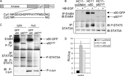
Figure 8.
Expression of s80Cyt2 induces STAT5A activity in HC11 cells. (A) Schematic diagram of s80HER4 (i.e., the intracellular domain of HER4) indicating the two naturally occurring cytoplasmic splice variants of human ErbB4/HER4. (B) Western analyses of ErbB4 or STAT5A immunoprecipitates from cells cultured overnight in serum-free media and then treated ± HB-EGF for 30 min. ErbB4 immunoprecipitates (made with a polyclonal ErbB4 antibody) were analyzed by Western with a monoclonal ErbB4 antibody. Stat5A immunoprecipitates were analyzed by Western with antibodies against phospho-STAT5 or STAT5A. (C) HC11-s80 and HC11-s80Cyt2 cells were separated into cytoplasmic and nuclear lysates, which were used for immunoprecipitation (described below) or directly for Western with an antibody against phospho-c-jun. ErbB4 immunoprecipitates were analyzed by Western analysis for phospho-tyrosine; STAT5A immunoprecipitates were analyzed by Western for phospho-STAT5. (D) Cells (indicated in legend) were transiently transfected with pβCasein-lux. Cells were maintained in serum-free media for the final 24 h of culture. Luciferase activity was measured as described above. n = 3.

