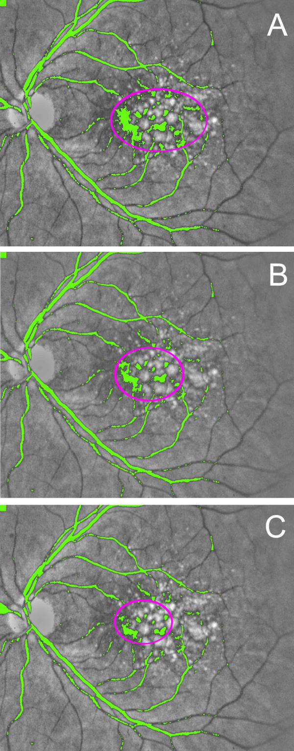
Figure 1.
Iterative macular background leveling. Processing takes place in the green channel; gray scale is used here for better reproduction. (A) All pixels darker than a fixed threshold are marked in a pseudo-color, in this case green. Note that green areas consist of the darker points in the central background, and the retinal vessels. The operator selects the size of the magenta oval such that it is just large enough to include the darker points in the central background, ignoring the retinal vessels. All pixels within this oval (both background and drusen) are brightened by 2 color intensity scale units. (B) The image created in (A) is subsequently fed back into the same algorithm. Note that all pixels darker than the same threshold are again marked in green, but the central region of darkness becomes smaller and is enclosed by a smaller magenta oval. Visualization of the retinal vessels is unaffected. The new region within the oval is further brightened as before. (C) The image created in (B) is sent back through the same algorithm. Note that the central darker region is again reduced in size, since points at the edges that were just below the threshold in (B) have been brightened. The process is continued until the macular background is uniform.

