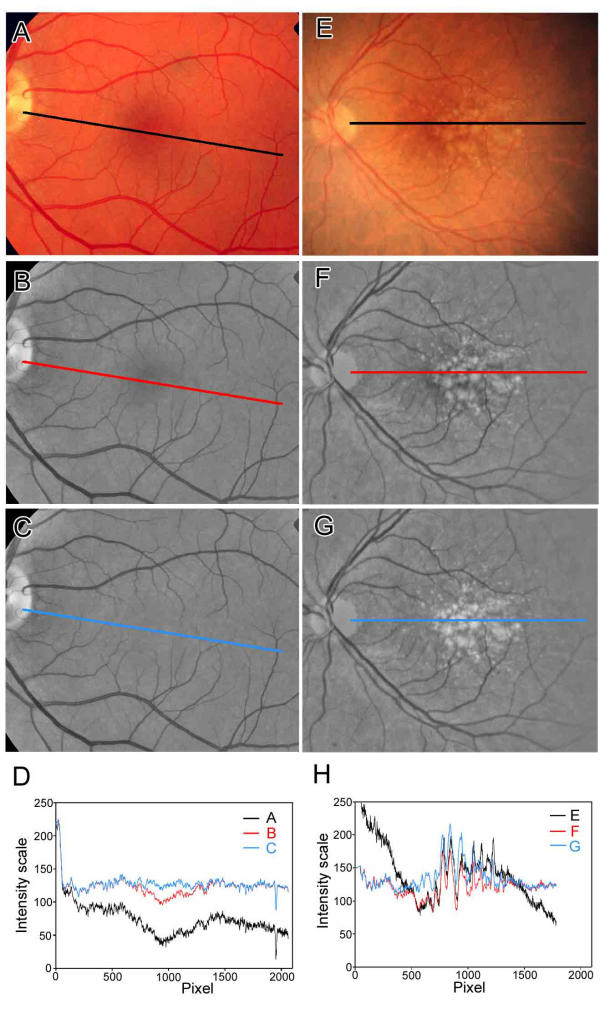
Figure 2.
Line scans demonstrating macular background leveling for drusen recognition. Left Panel. The line scans through a normal fundus image (A-D) demonstrate the effects of large-scale shading correction and macular background leveling. The color image (A) was processed and scanned in the green channel (B-D). For purposes of illustrating detail, these images were processed at higher resolution (1000 pixels from central macula to disc edge). The width of the scan line was 10 pixels. The original image (normal fundus A) is darker in the temporal quadrant and this is illustrated by the line scan (black line in D). The latter darkening is eliminated in the shading-corrected image (B) and verified by the line scan (red line in D). Note that the red line for the shade-corrected image still has a central dip of about 25 color intensity scale units corresponding to the darker central macula. This central dip is corrected with background leveling. The macular background is now quite uniform and the center is no longer dark (C). This is shown by the line scan (blue line in D). Right Panel. The line scans through a drusen-containing image (E-H) also demonstrate the effects of large-scale shading correction and macular background leveling. The color image (E) was processed and scanned in the green channel (F-H). The original image (drusen fundus E) is darker in the temporal quadrant and this is illustrated by the line scan (black line in H). The latter darkening is eliminated in the shading-corrected image (F) and verified by the line scan (red line in H). Note that the red line for the shade-corrected image (panel H, red line) still has a central dip of about 25 color intensity scale units corresponding to the darker central macula between pixels 500 and 750, but this is harder to interpret due to the multiple jagged drusen peaks. This central dip is corrected with background leveling (blue line in H). In the fundus image, the macular background between the drusen is also now uniform (G) without central darkness. Note that this technique also raises the brightness of associated drusen (G) along with the background. This is verified by the line scans, in which the drusen peaks in the central macula of the background-leveled image (blue peaks in H) are raised above the peaks in the image before background leveling (red peaks in H), allowing a uniform threshold.

