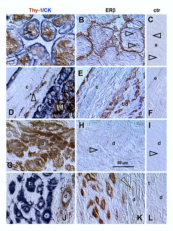
Figure 1.
Dual color for Thy-1 differentiation protein and cytokeratin (Thy-1/CK), single color ERβ, and control staining (ctr) in CV and placental membranes. [A–C] Corresponding sections through the terminal villi of normal term placenta: s, syncytiotrophoblast; arrowheads, villous blood sinusoids. [D–F] Placental membranes: e, amniotic epithelium; c, connective tissue; t, extravillous trophoblast; t/d, trophoblast/decidua interface; d, decidual cells; arrowhead, Thy-1+ fibroblasts. [G–I] Placental basal plate decidua (d); arrowheads, comparison of ERβ and control staining. [J–L] Placental basal plate extravillous trophoblast (t) and decidua compartments (d); arrowhead indicates nuclear staining of decidual cell. No hematoxylin counterstain. Details in text.

