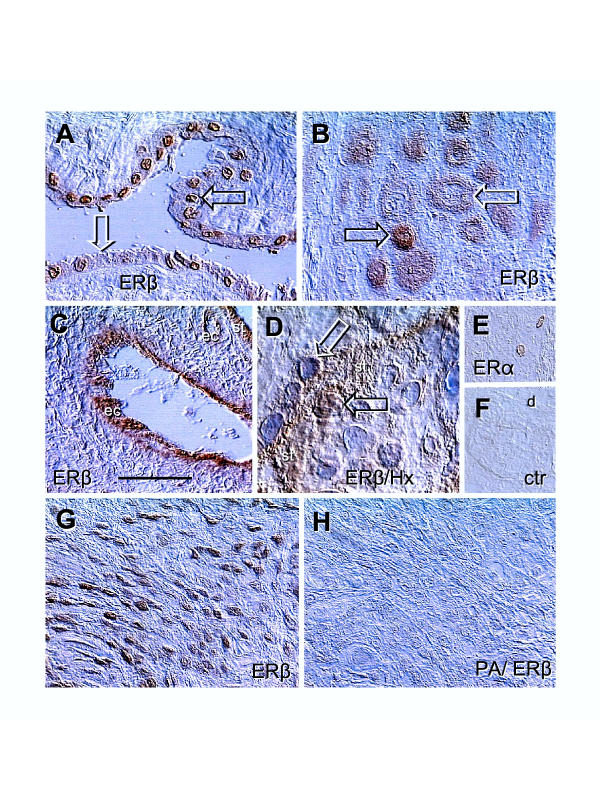
Figure 2.
Immunolocalization of ERs in placental membranes and ERβ peptide-absorbed antibody control. [A] Amniotic epithelium with ERβ+ (solid arrow) and ERβ- nuclei (white arrow). [B] Basal plate EVT with high (solid arrow) and diminishing ERβ staining (white arrow). [C] Immunolocalization of ERβ in vascular endothelial cells (ec) of a stem villus. [D] Cytotrophoblast cell merging with ST (st) shows nuclear ERβ immunoreactivity (solid arrow) but ST nuclei are unstained (white arrow). [E] Rarely seen nuclear ERα immunoreactivity in the basal plate decidual cells. [F] Control. [G] EVT in lower magnification, representing ERβ positive control for [H], which is a parallel section incubated with peptide-absorbed ERβ antibody (PA/ERβ). Bar in C = 50 μm for [A–C], 20 μm for [D], and 100 μm for [E-H]. No nuclear counterstain except in panel [D]. Details in text.

