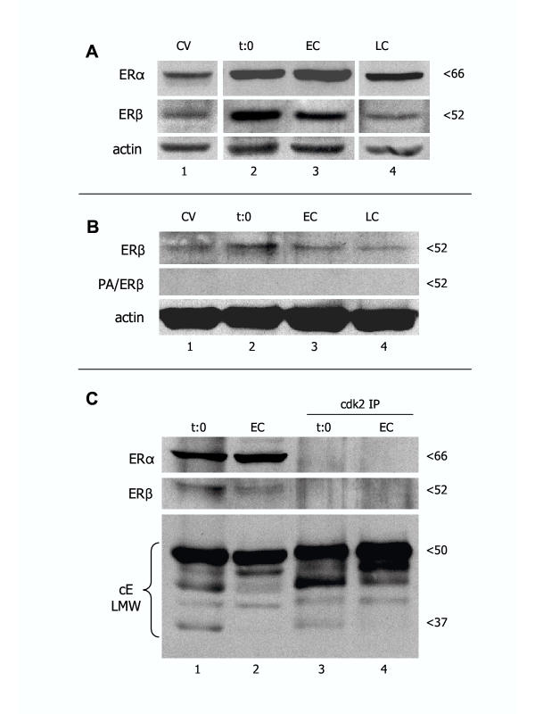
Figure 6.
Representative western blot analysis of ERα and ERβ expression in CV, time 0 separated trophoblast cells (t:0), and early (EC) and late trophoblast cultures (LC). [A] All lanes are from the same normal placenta and corresponding cultures, and from the same blot. [B] Absorption of ERβ antibody with blocking peptide (PA/ERβ) resulted in a lack of ERβ band detection in CV from another placenta and derived trophoblast cultures. [C] Demonstration of ERα and ERβ expression in original protein lysates (lanes 1 and 2) and lack of reactivity with cdk2 immunoprecipitates (cdk2 IP; lanes 3 and 4) from time 0 and early trophoblast cultures. Bottom is the positive IP control showing cyclin E low molecular weight (cE LMW) protein variants in both original lysates and cdk2 IPs.

