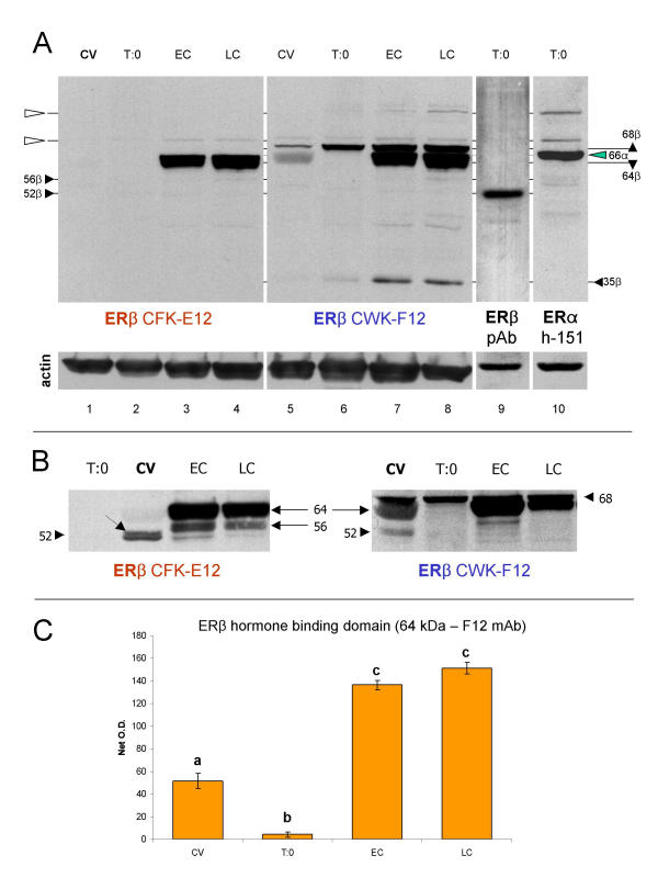
Figure 8.
Western blot analysis of ERβ hormone binding domain with E12 and F12 monoclonal antibodies. [A] Both antibodies identify ~64 kDa band, abundant in early (EC) and late cultures (LC). The F12 antibody (lanes 5–8) also identifies a distinct ~68 kDa and ~35 kDa bands, which show enhanced expression in trophoblast cultures. Note a virtual lack of ~52 kDa ERβ band in lanes 1–8, identified by polyclonal antibody [ERβ pAb (N-19)] in lane 9. The N-19 antibody (lane 9) does not show any reactivity except at ~52 kDa. Lane 10 shows ERα (h-151 antibody) band at 66 kDa (green arrowhead), which does not interfere with ~64 and 68 kDa reactivity of E12 and F12 antibodies. Open arrowheads indicate narrow bands, possibly non-specific reactivities shared among monoclonal antibodies. [B] Protein extracts from another placenta show similar character of expression of ~64 kDa ERβ hormone binding domain. Relatively strong ~56 kDa E12 immunoreactivity is also evident in trophoblast cultures. Both antibodies identify a distinct ~52 kDa ERβ in protein extracts from chorionic villi (CV), which is barely detectable in trophoblast cultures. Dotted arrow indicates adjacent ~53 kDa band. [C] Quantitative evaluation of ~64 kDa ERβ protein identified by F12 antibody in [A]. Each column represents a mean from six net O.D. measurements ± SD. Different column superscripts indicate P < 0.01.

