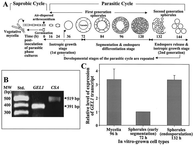
FIG. 3.
(A) Diagrammatic representation of the saprobic and parasitic cycles of C. posadasii. The developmental stages of first- and second-generation parasitic cells are identified by incubation times after inoculation of parasitic cultures, as previously reported (17). (B) RT-PCR amplification of GEL1 and CSA genes expressed in C. posadasii-infected, murine lung tissue. Fluorescence microscopy of Blankofluor-stained whole mounts of infected tissue revealed large numbers of spherules in the endosporulation stage of development (data not shown). The RT-PCR results are representative of three separate preparations of total RNA isolated from three infected mice. (C) QRT-PCR of C. posadasii GEL1 expression at different stages of saprobic and parasitic cell growth in vitro. The relative amounts of GEL1 transcript in the mycelial and spherules (132-h endosporulation stage) were compared to the transcript level in spherules at early segmentation stage (72 h), which was given an arbitrary value of 1. The data indicate that a significant increase in expression of the GEL1 gene occurred during the endosporulation stage of the parasitic cycle.

