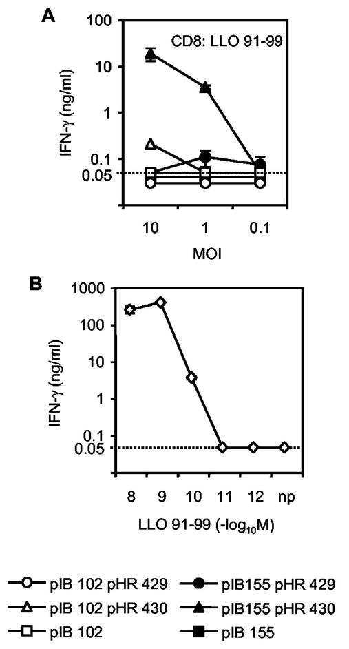
FIG. 3.
Effect of Yersinia-mediated hypertranslocation on MHC class I-restricted antigen display. Presentation of the MHC class I-restricted epitope LLO91-99 was measured in an in vitro antigen presentation assay with an epitope-specific CD8 T-cell line. (A) APC were infected with Y. pseudotuberculosis pIB102(pHR429) or pIB155(pHR429), each of which secretes the chimeric YopE/LLO protein, or with pIB102(pHR430) or pIB155(pHR430), which translocates or hypertranslocates the YopE/LLO fusion protein, respectively. Nontransformed pIB102 and pIB155 were used as controls. Cells were infected at an MOI of ∼10, ∼1, or ∼0.1. (B) The sensitivity of the CD8 T-cell line was monitored after loading of APC with graded amounts of LLO91-99 peptide. Activation of T cells was measured as the amount of IFN-γ secreted into the culture supernatant. The means and standard deviations of duplicate cultures are indicated. The dotted line at 0.05 ng/ml indicates the detection limit of the IFN-γ ELISA.

