Figure 4.
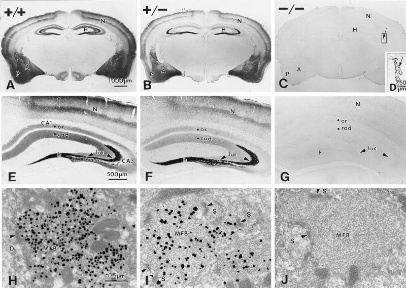
Timm stain is undetectable in the brains of ZnT3−/− mice. Comparison of Timm stain between brains of ZnT3+/+ (A, E, and H), ZnT-3+/− (B, F, and I), and ZnT-3−/− mice (C, G, and J). (A–C) Coronal sections through the midbrain. Timm stain in the hippocampus (H), piriform cortex (P), neocortex (N), and amygdala (A) was conspicuous in the ZnT3+/+ brain (A), reduced in the ZnT3+/− brain (B), and undetectable in the brains of ZnT3−/− mice (C). (D) Higher magnification of the choroid plexus from the lateral ventricle (indicated area in C). Timm stain was unperturbed in the ZnT3−/− choroid plexus. (E–G) Higher magnification of Timm-stained hippocampi from ZnT3+/+ (E), ZnT3+/− (F), and ZnT3−/− (G) mice. Timm stain was reduced (ZnT3+/−) or absent (ZnT3−/−) in the hilus (h), s lucidum (luc) of CA3, and s oriens (or) and s radiatum (rad) of CA1 and CA3. (H–J) Electron micrographs of Timm-stained MFBs in s lucidum of CA3, taken from a ZnT3+/+ (H), a ZnT3+/− (I), and a ZnT3−/− (J) mouse. Timm-positive vesicles were abundant in ZnT3+/+ MFBs, whereas fewer vesicles were Timm-positive in ZnT3+/− MFBs, and no Timm-positive vesicles were present in ZnT3−/− MFBs. Arrowheads represent synaptic contacts made with a dendrite (D) and dendritic spines (S).

