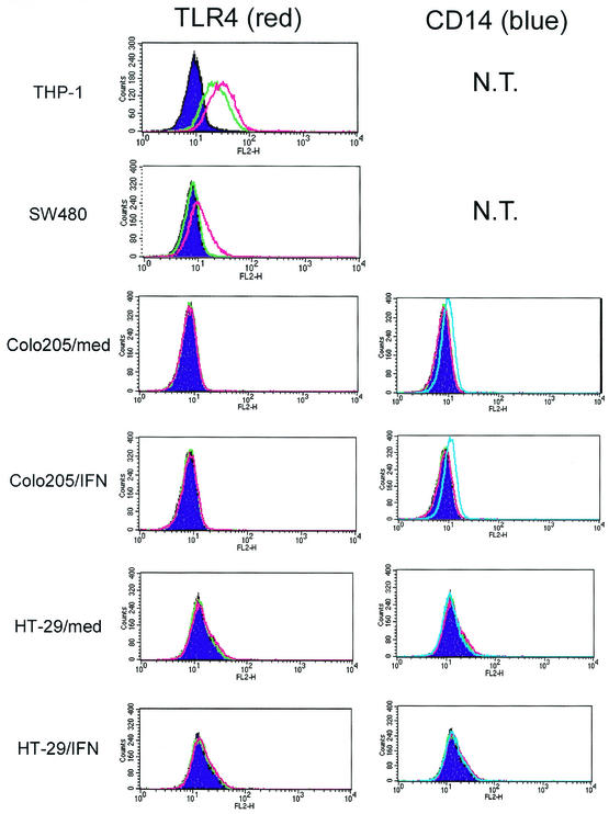
FIG. 5.
Expression of TLR4 and CD14 by intestinal epithelial cells. Cells were cultured with IFN-γ (10 ng/ml) or medium for 16 h, collected, stained with PE-labeled anti-TLR4 MAb (red line), PE-labeled anti-CD14 MAb (blue line), or PE-labeled isotype control (green line), and analyzed by FACS as described in Materials and Methods. Filled purple areas show nonstaining control. Left panels show TLR4 staining, and right panels show CD14 staining. In all epithelial cell lines, significant background staining with isotype control could not be detected. However, significant background staining with isotype control MAb (green line) could be detected in THP-1 cells. This background staining seems to be mediated by Fc receptors expressed on THP-1 cells.

