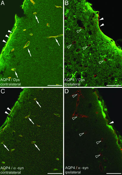
Fig. 1.
Immunofluorescence analysis of brains subjected to MCAO (neocortex; 24 hr of reperfusion). (A and C) Contralateral neocortex, area opposite to ischemic core. (B and D) Central part of ischemic cortex (Fig. 5, region 4). Yellow labeling surrounding vessels in contralateral cortex indicates colocalization (arrows) of AQP4 (green) and dystrophin (red in A) and α-syntrophin (red in C). In contrast, vessels in the ischemic cortex are associated with a red signal (open arrowheads), indicating the absence of AQP4 and retention of dystrophin (B) and α-syntrophin (D). Filled arrowheads indicate the subpial endfeet. Dys, dystrophin; α-syn, α-syntrophin. (Scale bar: 20 μm.)

