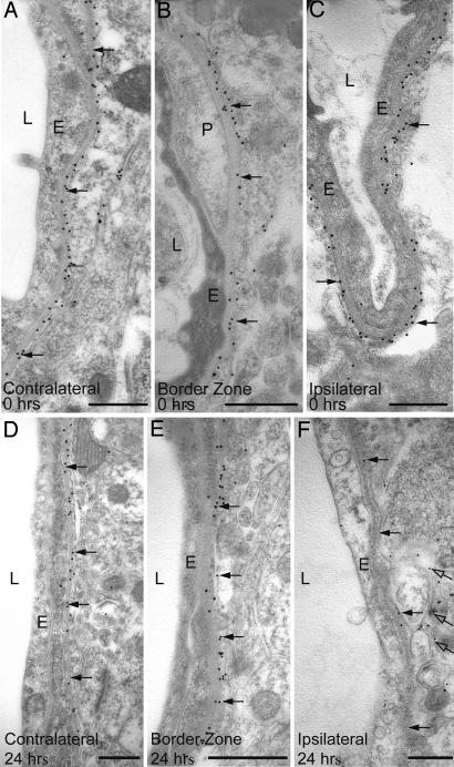
Fig. 2.
Immunogold analysis of AQP4 expression immediately (0 hr in A–C) and 24 hr (D–F) after the onset of reperfusion (0 hr). At 24 hr, there is a pronounced reduction in the number of gold particles (arrows) over perivascular membranes in central part of the ischemic cortex (F) compared with the border zone (E) and contralateral side (D). In the ischemic cortex, AQP4 labeling remains over the abluminal membrane of the perivascular endfeet (open arrows in F). E, endothelial cells; L, vessel lumen; P, pericyte. (Scale bar: 0.5 μm.)

