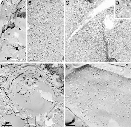
Fig. 4.
Conventional freeze fracture images from contralateral cortex (A and B), cortical border zone (C and D), and central part of ischemic lesion (E and F), all from 24 hr of reperfusion after MCAO. (A) Low-magnification image from contact region between astrocyte endfoot (white asterisk) and the edge of capillary (black asterisk in cytoplasm). Boxed area encloses P-face of astrocyte, shown at higher magnification in B. Nu, endothelial nucleus. (B) High-magnification view of P-face of astrocyte endfoot plasma membrane, showing >100 AQP4 arrays in ≈0.3 μm2. (C) P-face of astrocyte endfoot in border zone has AQP4 arrays at approximately the same density as in contralateral control. (D) E-face view of AQP4 imprints, also from border zone. (E) Low-magnification view of capillary in central part of ischemic lesion. Membrane debris is present in the lumen of the capillary. Boxed area including presumptive astrocyte endfoot shown at higher magnification in F. The black asterisk indicates endothelial cytoplasm. (F) High-magnification image of E-face of presumptive astrocyte endfoot in central part of ischemic lesion. AQP4 arrays were not detected in this or any other membrane adjacent to capillaries. The black asterisk indicates endothelial cytoplasm. B–D and F are at the same magnification. (Calibration bars: A and E, 1 μm; B, C, and F, 0.1 μm.)

