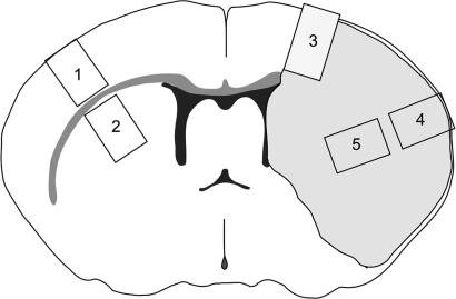
Fig. 5.
Diagram of coronal slice through the forebrain. The shaded area on the right side is the area affected by ischemia. Specimens were dissected from the following: 1, contralateral neocortex (control); 2, contralateral striatum (control); 3, cortical border zone; 4, central part of ischemic cortex; and 5, striatal part of the ischemic core.

