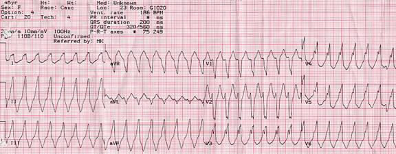
Figure 1.

Wide complex tachycardia in a 45 year old woman with a structurally normal heart. This rhythm was incidentally noted on a cardiac monitor during an elective surgery. The patient had a similar episode 4 years earlier in the context of another elective surgery. She was completely asymptomatic during this rhythm.
Note the left bundle, right axis morphology of the QRS complex. The width of the QRS complex is 200 milliseconds. The initial r wave in V1 is about 35 milliseconds in duration. There is a notch on the down stroke of the S wave in V1. All these morphologic features point to a diagnosis of VT. There is no precordial concordance in ECG leads V1 through V6 however, a feature more frequently seen in SVTs. Adenosine (12 mg IV) failed to terminate the tachycardia or change the rhythm. This WCT represents an idiopathic VT originating from the right ventricular outflow tract. Paper speed is 25 mm/s.
