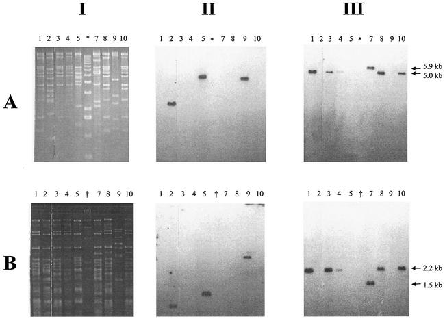
FIG. 2.
RFLP and Southern blot analysis of phage genomic DNA. (A) EcoRI-digested DNA; (B) HincII-digested DNA. Columns: I, RFLP patterns on TAE-agarose gel; II, Southern blot analysis of I with a Stx1-specific probe; III, Southern blot analysis of I with an Stx2-specific probe. Phage DNA were from the following sources: lane 1, φE86654-Stx2; lane 2, φE86654-Stx1; lane 3, φE85539-Stx2; lane 4, φE85539-Stx2′; lane 5, φE83819-Stx1; lane 7, φE83819-Stx2; lane 8, φE45040-Stx2; lane 9, φD155-Stx1; lane 10, φPS14-Stx2; lane ✽, molecular mass marker X (Roche); lane †, molecular weight marker II (Roche). Sizes: ✽, 12,216, 1,1198, 10,180, 9,162, 8,144, 7,126, 6,108, 5,090, 4,072, 3,054, 2,036, 1,636, 1,018, 517, 506, 396, 433, 298, 220, 201, 154, 134, and 75 bp; †, 23,130, 9,416, 6,557, 4,361, 2,322, 2,027, 564, and 125 bp.

