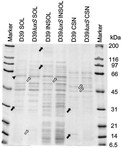
FIG. 4.
Protein profiles of subcellular fractions of D39 and D39luxS. Supernatants (100,000 × g) (SOL) and pellets (INSOL) of French pressure cell lysates of S. pneumoniae cells, as well as trichloroacetic acid-precipitated culture supernatant from the original THY cultures (CSN), were separated by SDS-PAGE (12% gel) and stained with Coomassie blue, as described in Materials and Methods. Solid arrows indicate protein species present in D39 but absent in D39luxS. Open arrows indicate protein species present in D39luxS but absent in D39. The arrowhead indicates a protein band with altered mobility between D39 and D39luxS. Molecular masses of size markers are indicated on the right of the figure.

