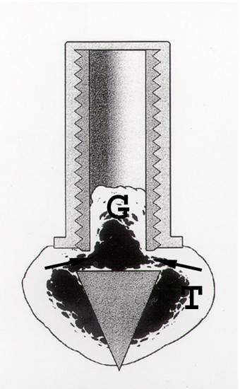
Figure 1.

The bone conduction chamber in situ in the proximal tibia (T). The graft (G) is placed in the chamber and mesenchymal tissue grows in from the bottom upwards into the bone graft, which subsequently remodels. Arrows point at ingrowth openings. (Reproduced with permission from Eur J Exp Musculoskel Res 2: 70, 1993).
