FIGURE 5.
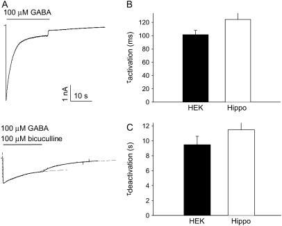
Current decay during GABA application is observed in hippocampal neurons expressing the ρ1 subunit. (A) Current traces of GABA (100 μM)–evoked responses in a hippocampal neuron transduced with an adenovirus expressing the ρ1-subunit without (top) or with (bottom) coapplication of 100 μM bicuculline. The dashed lines are the best-fitting monoexponential function fit to the activation and the deactivation phases of the bicuculline-resistant current. See Cheng et al. (7) for further details on the ρ1 subunit-expressing adenovirus. (B) A bar diagram summary of the approximate monoexponential time constant of activation (top) and deactivation (bottom) for HEK293 cells (n = 12) and hippocampal neurons (n = 37); p = 0.178 for activation and p = 0.192 for deactivation time constants based on a two-tailed t-test; thus, the difference in mean was not statistically significant for either measure.

