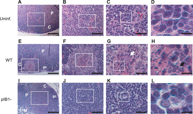
Figure 2. MLN Histopathology during WT or pIB1− Infection.
BALB/c mice were intragastrically inoculated with 2 × 10⁹ WT or IP2666 pIB1−, and MLN were processed for H&E staining 4 d later. MLN sections from uninfected (Uninf.) (A–D), WT infected (E–H), and pIB1− infected (I–L) are shown at 15×, 40×, 90×, and 450× magnification. White boxes indicate magnified areas in the next slide. White arrow points to the bacterial foci, black arrow to neutrophils. Scale bars correspond to 133 μm for 15× magnification, 50 μm for 40× magnification, 22 μm for 90× magnification, and 4.4 μm for 450× magnification. Pictures shown are representative of multiple fields and samples from 16 MLN infected with WT or 14 MLN infected with pIB1−.
C, cortex; gc, germinal center; M, medulla; P, paracortex.

